File:Ultrasound of gallbladder adenomyosis.jpg

Page contents not supported in other languages.
This is a file from the Wikimedia Commons
From Wikipedia, the free encyclopedia

Ultrasound_of_gallbladder_adenomyosis.jpg(472 × 554 pixels, file size: 109 KB, MIME type: image/jpeg)

Summary

Description
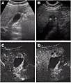
English: Non-contrast abdominal ultrasound and contrast-enhanced ultrasound (CEUS) of adenomyomatosis of the gallbladder.
a The fundus of the gallbladder wall was thickened and the GB wall was obscure.
b The intramural echogenic foci were detected by high frequency transducer.
c CEUS—arterial phase (22 s) —heterogeneous hyper-enhancement and wall was intact.
d CEUS—venous phase (34 s) the anechoic spaces were more clear
Date
Source (2015). "Contrast-enhanced ultrasonography diagnosis of fundal localized type of gallbladder adenomyomatosis". BMC Gastroenterology 15 (1). DOI:10.1186/s12876-015-0326-y. ISSN 1471-230X.
Author ShaoShan Tang, LiPing Huang, Yao Wang and YiJiao Wang

Licensing

w:en:Creative Commons
attribution
This file is licensed under the Creative Commons Attribution 4.0 International license.
You are free:
  • to share – to copy, distribute and transmit the work
  • to remix – to adapt the work
Under the following conditions:
  • attribution – You must give appropriate credit, provide a link to the license, and indicate if changes were made. You may do so in any reasonable manner, but not in any way that suggests the licensor endorses you or your use.

Captions

Add a one-line explanation of what this file represents

Items portrayed in this file

depicts

27 July 2015

File history

Click on a date/time to view the file as it appeared at that time.

Date/TimeThumbnailDimensionsUserComment
current13:28, 1 February 2018Thumbnail for version as of 13:28, 1 February 2018472 × 554 (109 KB)Mikael HäggströmUser created page with UploadWizard
The following pages on the English Wikipedia use this file (pages on other projects are not listed):

Global file usage

The following other wikis use this file: