User:Doctor Cornea/sandbox
Eye aging[edit]
Eye aging is the natural changes that occur in the eye as people age. The eyes contain various highly specialised tissues that work together to detect light and provide vision. As people get older, numerous tissues in the eye undergo physiological changes that may lead to vision problems, including vision loss and other eye diseases. While the specific causes and prevalence of vision loss differ across regions, it generally increases with age. There are a range of eye diseases related to eye aging, but there are also ways to delay the aging process, such as keeping a healthy lifestyle and having regular eye exams. Future research in this area may provide new strategies for promoting healthy aging of the eyes.
Function and anatomy of the eye[edit]
Highly specialised tissues are spread throughout the eyes. They are fundamental to maintaining the complex vision-generating process machinery. There are two major refractive surfaces focusing the light rays entering the front eyes, which are the cornea and the lens. The cornea contributes about two-thirds of the refractive power while the lens contributes the remaining[1]. Then, the light rays pass through the vitreous humor reaching the retina, where densely populated first light-sensitive neurons (rod and cone photoreceptors) are located.

The retina located at the back of the eye is an organised structure, and it has an indispensable role in the transduction and transmission of signals. When the photoreceptors are stimulated by lights, neural signals are generated and passed through the second neurons (bipolar cells) and tertiary neurons (retinal ganglion cells).[2] Eventually, the signals coalesced into the optic nerve are sent to the visual cortex in the brain for interpretation.
Physiological changes and causes[edit]
Physiological changes in all ocular tissues occur along with age. The progressive change of structure will devastatingly lower its efficiency, or even lead to the malfunction of tissues.

Cornea, which is a powerful refractive surface, will flatten, causing an attrition of endothelial cells. The corneal endothelial cells are responsible for maintaining the cornea’s structural integrity.[2] However, with aging, there is a decrease in corneal endothelial cell density. Approximately 2000 cells/mm2 of corneal endothelial cell density are decreased from birth to older age.[3] Not only the cornea, but the shape of the trabecular meshwork also alters. Research tracking specimens from newborns to 81 years old found that there are progressive decreases in trabecular endothelial cell density and absolute cell number caused by aging.[4] This phenomenon leads to a change in the conformation of the trabecular meshwork. The configuration transforms from a wedge shape into a shorter and rhomboidal form.[5][6]
Moreover, the ciliary body becomes collagenized as time passes. The ciliary body contains ciliary muscles that could change the thickness and curvature of the lens. As the cellularity of the ciliary body diminishes with age, they appear to be shorter and fail to complete their function.[3] In addition, the aging of the crystalline lens will occur. Its shape will be changed from a reinform configuration to a more oval shape. Apart from the stiffening of the lens with advancing age, the crystallins protein in the lens gradually loses its transparency. This is possibly due to the increasing oxidation stress along with age.[2]
The cells in the retina are continuously exposed to light, which makes them susceptible to damage from light. In addition, the reduction of nuclei in the outer nuclear layer of the retina may occur with age.[3] Various problems, including the decline in visual acuity and colour perception threshold, are inevitably arising with age. Research has also shown that rods degenerate before cones in the macula as time passes. Therefore, rod-mediated (scotopic) sensitivity has a larger decline than cone-mediated (photopic) sensitivity. Hence, the dysfunction of the rod slows down the dark adaptation.[7]
Furthermore, connective tissues within the optic nerve become more abundant with age, affecting the exchange of materials between the capillaries and nerve fibres. This results in the accumulation of cellular and extracellular materials in the optic nerve fibre bundles.[8] Such accumulation inhibits the transmission of nerve signals to the brain, hindering the generation of vision.
Prevalence[edit]
The prevalence of vision impairment tends to increase with age. In China, the distribution of vision impairment has changed significantly between 1999 and 2019. During this period, the number of people with moderate vision impairment increased by 67%, while those with severe vision impairment showed a 14% increase, and those with blindness experienced a 35% increase[9].
Related diseases[edit]
Age-related eye diseases are conditions that affect the structure and function of the eyes as people age. These conditions can lead to loss of vision and may require medical treatment. They can have a significant impact on a person's quality of life. Common age-related eye diseases include age-related macular degeneration (AMD), cataracts, glaucoma and dry eye syndrome.[10]

[edit]
Age-related macular degeneration (AMD) is an eye disorder in which the macula, a part of the retina responsible for central vision, progressively deteriorates. This can result in a loss of central vision, making it difficult for people to read, see fine details, and recognize faces. There are two major types of AMD: dry AMD, which progresses relatively slower, and wet AMD, which can lead to rapid vision loss.[11]
Cataracts[edit]
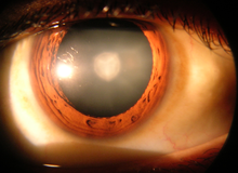
Cataract is another common age-related eye disease that occurs when the lens of the eye becomes opaque or cloudy. This can cause the vision to become hazy, blurry, or distorted. Cataracts can develop gradually over time and may require surgical treatment.[12]

Glaucoma[edit]

Glaucoma is a common eye condition where the optic nerve is damaged.[13] The optic nerve is responsible for transmitting visual information from the eye to the brain. In the early stages, glaucoma may not show noticeable symptoms. However, as the condition progresses, it can cause loss of vision, usually starting with peripheral vision, and possibly leading to total blindness if the condition is left untreated.[14]
Dry eye syndrome[edit]
Dry eye syndrome is an eye condition in which the eyes cannot produce enough tears, or when the tears produced evaporate too quickly.[15] This can lead to discomfort in the eyes, including itching, redness, and sensitivity to light. It can also result in blurred or distorted vision.[16] Common treatments for dry eye syndrome include eye drops, antibiotics, changes in lifestyle and other therapies, such as intense pulsed light therapy.[17]
Early detection and treatment of these age-related eye diseases are important to preserve vision and eye health. It is recommended to have regular eye exams to monitor for these conditions and address any potential eye concerns.
Delay and prevention[edit]
There are a range of ways to delay eye aging and reduce the risk of eye diseases.
Healthy lifestyle habits, which include having a balanced diet, having regular exercise and avoiding smoking can help delay eye aging. These habits also help prevent health conditions such as diabetes and high blood pressure, which may contribute to eye diseases.
Protecting the eyes from harmful ultraviolet (UV) radiation can help delay the aging of the eyes. By wearing sunglasses that block out UV radiation, the eyes can be protected from the damage caused by the harmful rays from the sun. Wearing protective eyewear when taking part in activities which may expose the eyes to strong lights can also help preserve the eyes.
Regular eye exams are effective in detecting any change in the eyes early on. They are essential in maintaining eye health. The exams can identify the various age-related eye diseases before their progression and the resulting significant loss of vision.[18][19]
Eye exercise and relaxation techniques can help delay eye aging. The exercise improves the strength and flexibility of eye muscle, reduces eye strain and fatigue, and improves visual acuity (the ability of the eye to distinguish the shapes and the details of objects).
Research and future development[edit]
Future research directions in the field of eye aging include the development of new diagnostic tools and technology for the early detection of age-related eye diseases, the development of new drugs, and the research in novel therapies, such as gene therapy and stem cell therapy.[20]
Research can also focus on the impact of different lifestyle factors, such as diets and exercise, on the condition of eye aging, to develop more strategies for promoting healthy aging of the eye.[21]
References[edit]
- ^ Kaplan, Henry J. (2007), "Anatomy and Function of the Eye", Immune Response and the Eye, Basel: KARGER, pp. 4–10, retrieved 2023-04-06
- ^ a b c Lin, Jonathan B; Tsubota, Kazuo; Apte, Rajendra S (2016-03-10). "A glimpse at the aging eye". npj Aging and Mechanisms of Disease. 2 (1). doi:10.1038/npjamd.2016.3. ISSN 2056-3973.
- ^ a b c Grossniklaus, Hans E.; Nickerson, John M.; Edelhauser, Henry F.; Bergman, Louise A. M. K.; Berglin, Lennart (2013-12-13). "Anatomic Alterations in Aging and Age-Related Diseases of the Eye". Investigative Opthalmology & Visual Science. 54 (14): ORSF23. doi:10.1167/iovs.13-12711. ISSN 1552-5783.
- ^ Alvarado, Jorge; Murphy, Collin; Juster, Richard (1984-06). "Trabecular Meshwork Cellularity in Primary Open-angle Glaucoma and Nonglaucomatous Normals". Ophthalmology. 91 (6): 564–579. doi:10.1016/s0161-6420(84)34248-8. ISSN 0161-6420.
{{cite journal}}: Check date values in:|date=(help) - ^ McMenamin, Paul G.; Lee, William R.; Aitken, Dorothy A.N. (1986-02). "Age-related Changes in the Human Outflow Apparatus". Ophthalmology. 93 (2): 194–209. doi:10.1016/s0161-6420(86)33762-x. ISSN 0161-6420.
{{cite journal}}: Check date values in:|date=(help) - ^ Crouch, Devon J.; Sheridan, Carl M.; D'Sa, Raechelle A.; Willoughby, Colin E.; Bosworth, Lucy A. (2021-03). "Exploiting biomaterial approaches to manufacture an artificial trabecular meshwork: A progress report". Biomaterials and Biosystems. 1: 100011. doi:10.1016/j.bbiosy.2021.100011. ISSN 2666-5344.
{{cite journal}}: Check date values in:|date=(help) - ^ Jackson, Gregory R; Owsley, Cynthia; Curcio, Christine A (2002-06). "Photoreceptor degeneration and dysfunction in aging and age-related maculopathy". Ageing Research Reviews. 1 (3): 381–396. doi:10.1016/S1568-1637(02)00007-7.
{{cite journal}}: Check date values in:|date=(help) - ^ "Ophthalmic Pathology. An Atlas and Textbook. 2d Ed". Annals of Internal Medicine. 56 (6): 999. 1962-06-01. doi:10.7326/0003-4819-56-6-999_2. ISSN 0003-4819.
- ^ Xu, Tingling; Wang, Bingsong; Liu, Hua; Wang, Haidong; Yin, Peng; Dong, Wenlan; Li, Jianhong; Wang, Ya Xing; Yusufu, Mayinuer; Briant, Paul; Reinig, Nickolas; Ashbaugh, Charlie; Adelson, Jaimie; Vos, Theo; Bourne, Rupert (2020-12). "Prevalence and causes of vision loss in China from 1990 to 2019: findings from the Global Burden of Disease Study 2019". The Lancet Public Health. 5 (12): e682–e691. doi:10.1016/S2468-2667(20)30254-1.
{{cite journal}}: Check date values in:|date=(help) - ^ "Common Eye Disorders and Diseases | CDC". www.cdc.gov. 2022-12-14. Retrieved 2023-04-13.
- ^ National Eye Institude (22 June 2021). "Age-Related Macular Degeneration (AMD)". National Eye Institude.
- ^ National Eye Institude (13 January 2023). "Cataracts". National Eye Institude.
- ^ NHS (26 February 2021). "Glaucoma". The NHS website.
- ^ Tham, Yih-Chung; Li, Xiang; Wong, Tien Y.; Quigley, Harry A.; Aung, Tin; Cheng, Ching-Yu (2014-11). "Global prevalence of glaucoma and projections of glaucoma burden through 2040: a systematic review and meta-analysis". Ophthalmology. 121 (11): 2081–2090. doi:10.1016/j.ophtha.2014.05.013. ISSN 1549-4713. PMID 24974815.
{{cite journal}}: Check date values in:|date=(help) - ^ National Eye Institude (8 April 2022). "Dry Eye". National Eye Institude.
- ^ Craig, Jennifer P.; Nichols, Kelly K.; Akpek, Esen K.; Caffery, Barbara; Dua, Harminder S.; Joo, Choun-Ki; Liu, Zuguo; Nelson, J. Daniel; Nichols, Jason J.; Tsubota, Kazuo; Stapleton, Fiona (2017-07). "TFOS DEWS II Definition and Classification Report". The Ocular Surface. 15 (3): 276–283. doi:10.1016/j.jtos.2017.05.008. ISSN 1937-5913. PMID 28736335.
{{cite journal}}: Check date values in:|date=(help) - ^ Dell, Steven J (2017-12-31). "Intense pulsed light for evaporative dry eye disease". Clinical Ophthalmology. 11: 1167–1173. doi:10.2147/OPTH.S139894. ISSN 1177-5467. PMC 5488788. PMID 28790801.
{{cite journal}}: CS1 maint: PMC format (link) CS1 maint: unflagged free DOI (link) - ^ Yadav and Tandon (17 December 2019). "Comprehensive eye examination: what does it mean?". Community Eye Health. 32 (107) – via National Library of Medicine.
- ^ Shinohara, K.; Moriyama, M.; Shimada, N.; Nagaoka, N.; Ishibashi, T.; Tokoro, T.; Ohno-Matsui, K. (2013-11). "Analyses of shape of eyes and structure of optic nerves in eyes with tilted disc syndrome by swept-source optical coherence tomography and three-dimensional magnetic resonance imaging". Eye. 27 (11): 1233–1242. doi:10.1038/eye.2013.202. ISSN 1476-5454.
{{cite journal}}: Check date values in:|date=(help) - ^ Nazari, Hossein; Zhang, Li; Zhu, Danhong; Chader, Gerald J.; Falabella, Paulo; Stefanini, Francisco; Rowland, Teisha; Clegg, Dennis O.; Kashani, Amir H.; Hinton, David R.; Humayun, Mark S. (2015-09-01). "Stem cell based therapies for age-related macular degeneration: The promises and the challenges". Progress in Retinal and Eye Research. 48: 1–39. doi:10.1016/j.preteyeres.2015.06.004. ISSN 1350-9462.
- ^ Francisco, Sarah G.; Smith, Kelsey M.; Aragonès, Gemma; Whitcomb, Elizabeth A.; Weinberg, Jasper; Wang, Xuedi; Bejarano, Eloy; Taylor, Allen; Rowan, Sheldon (2020-09-18). "Dietary Patterns, Carbohydrates, and Age-Related Eye Diseases". Nutrients. 12 (9): 2862. doi:10.3390/nu12092862. ISSN 2072-6643.
{{cite journal}}: CS1 maint: unflagged free DOI (link)
